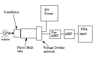
Equipment: NaI crystal mounted on a photomultiplier tube, preamplifier, amplifier, high-voltage power supply. Pulse-height analyzer (PHA), consisting of an analog-to-digital converter and histogramming memory. You will use the Tracor-Northern analyzer
Readings: Interactions of gamma-rays with matter, sect. 5.2; Scintillation counter, sect. 5.4.1, 5.4.2; Nuclear radiations and decay (consult a modern physics test, also study the decay schemes in Table 4.2); Radiation safety, sect. 4.5; Pulse-height analyser, sect. 4.3.3.
Key Concepts: Interactions of gamma-rays: photoelectric absorption, Compton scattering, pair production. Features of spectra: scatter peak, escape peak. PM tube operation.
2.1.1 NaI scintillator.
Measure spectra of a number of radioactive sources, for example 137Cs, 22Na, 60Co, 57Co, and 241Am, using the PHA. Transfer and save spectra for later analysis and plotting. Test for linearity between the central channel number of photopeak and corresponding known energies of the gammas.
2.1.2 Plastic scintillator.
If a plastic or liquid scintillator is available, measure spectra for several sources. Compare spectra obtained using plastic and NaI scintillators.
2.2.1 Features of pulse-height spectra. Interpret as fully as you can the spectrum of a monochromatic gamma-ray source like 137Cs. Be qualitative and quantitative. Then examine and interpret spectra for the other sources with, e.g., more than one gamma (60Co and 22Na), low-energy gammas (57Fe and 241Am).
2.2.2 Rest energy of the electron. From measured energies of Compton edges and the known energy of the gamma-rays, determine the rest energy of the Compton-scattered electron.
2.2.3 Photofraction. For several gammas, integrate the number of counts in the photopeak as well as the total number of counts detected from the corresponding gamma. The ratio is called the "photofraction". Determine how the photofraction depends on gamma energy and, if several crystals are available, crystal size.
2.2.4 Summing peaks. Record pulse-height spectra for weak sources in a "well" detector, if available, or on the face of a flat detector crystal in order to maximize the solid angle subtended by the detector. Search for summing peaks due to gammas that dump their energies into the crystal at the same time. Time "coincidences" can occur if gammas from different decays coincide accidentally in time or if gammas are emitted one after the other in the decay of a single nucleus ("accidental" and "true" coincidences). Accidental coincidences will be easier to observe for stronger sources. Test several weak and strong sources for true and accidental coincidences.
a. What is the efficiency of a detector? How could you measure it? Does it depend on the type and energy of the incident radiation, and how?
b. What is the energy resolution of a detector? How big is it for your NaI detectors? Does it depend on the energy of the gamma ray? Is there a fundamental lower limits on the energy resolution of a scintillation detector?
c. Suppose that two radiations arrive at the detector separated by a
very small time. What determines whether or not a summing peak will be
observed? Give an operational definition of the resolving time (or
time resolution) of your detector. Can you modify the time resolution?

Copyright Gary S. Collins, 1997-2002.