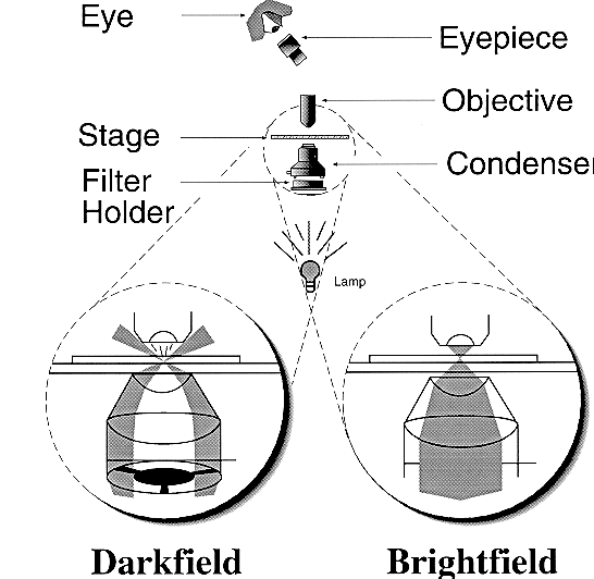
Figure 1.
This diagram compares the essential components of brightfield and darkfield microscopy. The difference in illumination (shown by stippling) of the sample between brightfield and darkfield is emphasized in the diagram. Darkfield utilizes a darkfield "stop" illustrated by the "spider stop" placed below the condenser. This stop blocks the center of the beam of light to produce a hollow cone of light. This light does not directly enter the objective lens. In contrast, a solid cone of light illuminates and enters the objective lens in brightfield. Only light that is scattered by the sample (depicted by the lines in the diagram) and enters the objective lens is seen as an image in darkfield.
Back to Darkfield Microscopy to Enhance Contrast
Back to Omoto's Bibliography