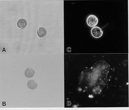
Figure 2.
Photomicrographs of Chlamydomonas are shown in Figure 2A-C and a buccal cell in Figure 2D. The difference in refractive indices of the media with the cell wall and other components of the algae are visualized in brightfield (Fig. 2A & B). Closing the condenser aperture diaphragm increases contrast allowing the flagella to be barely visible (Fig. 2A). Darkfield clearly shows many intracellular details as well as the flagella (Fig. 2C). Because these are unmounted living cells, they have moved a bit between the different exposures. Buccal cells are so similar in refractive index to the medium that photos of them could not be obtained in brightfield. Figure 2D shows buccal cells in darkfield; nucleus and other intracellular structures are clearly visible. Samples were photographed using CH2 student-grade Olympus microscope with 40X objective and 3.3X photo eyepiece on Kodak TMAX 400 film.
Back to Darkfield Microscopy to Enhance Contrast
Back to Omoto's Bibliography